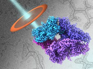
Atomic Resolution Cryo-EM Structure of B-galactosidase We report methods to account for radiation damage and local changes in defocus and image drift, enabling visualization of atomic resolution features in a cryo-EM density map of inhibitor-bound -galactosidase, and derivation of atom-specific measures of local flexibility of the bound inhibitor using constrained molecular dynamics simulations. Structure, 26(6), p. 848-85, 2018.
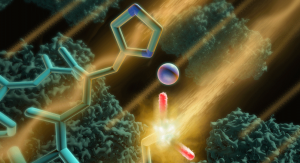
2.2 Å Resolution Cryo-EM Structure of β-galactosidase in Complex with a Cell-permeant Inhibitor Recent advances in cryo–electron microscopy allow structures of large macromolecules to be determined at near-atomic resolution. So far, though, resolutions approaching 2 Å, where features key to drug design are revealed, remain the territory of x-ray crystallography. Bartesaghi et al. achieved a resolution of 2.2 Å for a…
Structure of β-galactosidase at 3.2-Å Resolution Obtained by Cryo-Electron Microscopy The vast majority of high-resolution structures obtained using cryo-EM have been typically restricted to large, well-ordered entities such as helical or icosahedral assemblies or two-dimensional crystals. We show here that emerging methods in single-particle cryo-EM now allow structure determination at near-atomic resolution, even for much smaller protein complexes with…