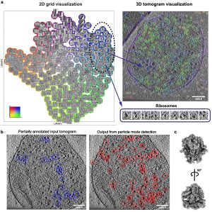
MiLoPYP: self-supervised molecular pattern mining and particle localization in situ Cryo-electron tomography (CET) allows the routine visualization of cellular landscapes in three dimensions at nanometer-range resolutions. When combined with single-particle tomography (SPT), it is possible to obtain near-atomic resolution structures of frequently occurring macromolecules within their native environment. Two outstanding challenges associated with CET/SPT are the automatic identification and…
Fab-dimerized glycan-reactive antibodies are a structural category of natural antibodies Natural antibodies (Abs) can target host glycans on the surface of pathogens. We studied the evolution of glycan-reactive B cells of rhesus macaques and humans using glycosylated HIV-1 envelope (Env) as a model antigen. We describe HIV-1 Env Fab-dimerized glycan (FDG)-reactive neutralizing Abs in HIV-1 vaccinated and simian-HIV (SHIV)-infected…
Single-particle cryo-EM structure of a voltage-activated potassium channel in lipid nanodiscs The structure of a voltage-activated potassium channel in lipid nanodiscs solved using cryo-electron microscopy is similar to previous X-ray structures, and provides insights into the mechanism of C-type inactivation. eLife, 7:e37558, 2018.
Cryo-EM structure of human rhodopsin bound to an inhibitory G protein For the first time, scientists have visualized the interaction between two critical components of the body’s vast cellular communication network, a discovery that could lead to more effective medications with fewer side effects for conditions ranging from migraine to cancer. The near-atomic resolution images obtained, show a G-protein coupled…
Cryo-electron Microscopy Structures of Chimeric Hemagglutinin Displayed on a Universal Influenza Vaccine Candidate Chimeric hemagglutinin proteins are set to undergo human clinical trials as a universal influenza vaccine candidate, yet no structural information for these proteins is available. Using cryo-electron tomography, we report the first three-dimensional (3D) visualization of chimeric hemagglutinin proteins displayed on the surface of the influenza…
Cryo-EM Structures of the Magnesium Channel CorA Reveal Symmetry Break upon Gating Magnesium ions (Mg2+) play essential roles in all living organisms. Bacteria and other prokaryotes rely upon the Mg2+-dependent channel CorA, which is composed of five identical subunits (A-E), to obtain these ions from their surroundings. Studies of CorA showed that, in contrast to most ligand-gated ion channels,…