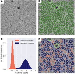
Automated systematic evaluation of cryo-EM specimens with SmartScope We present SmartScope, the first framework to streamline, standardize, and automate specimen evaluation in cryo-electron microscopy. SmartScope employs deep-learning-based object detection to identify and classify features suitable for imaging, allowing it to perform thorough specimen screening in a fully automated manner. A web interface provides remote control over the automated operation…
Fab-dimerized glycan-reactive antibodies are a structural category of natural antibodies Natural antibodies (Abs) can target host glycans on the surface of pathogens. We studied the evolution of glycan-reactive B cells of rhesus macaques and humans using glycosylated HIV-1 envelope (Env) as a model antigen. We describe HIV-1 Env Fab-dimerized glycan (FDG)-reactive neutralizing Abs in HIV-1 vaccinated and simian-HIV (SHIV)-infected…
Cryo-ZSSR: multiple-image super-resolution based on deep internal learning We present a multiple-image super-resolution (SR) algorithm based on deep internal learning designed specifically to work under low-SNR conditions typical of cryo-EM data. Our approach leverages the internal image statistics of cryo-EM movies and does not require training on ground-truth data. When applied to a single-particle dataset of apoferritin, we show…
Beam image-shift accelerated data acquisition for near-atomic resolution single-particle cryo-electron tomography To overcome the inherent low-throughput characteristic of CET data collection, improve the resolution of SVA and extend its application to a wider set of samples including low molecular weight targets, here, we: (1) use beam-image shift navigation to multiply the number of regions of interest imaged at each…
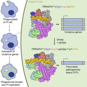
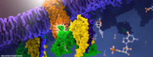
Structural Basis for Virulence Activation of Francisella tularensis The bacterium Francisella tularensis (Ft) is one of the most infectious agents known. Ft virulence is controlled by a unique combination of transcription regulators: the MglA-SspA heterodimer, PigR, and the stress signal, ppGpp. MglA-SspA assembles with the σ70-associated RNAP holoenzyme (RNAPσ70), forming a virulence-specialized polymerase. These factors activate Francisella pathogenicity island…
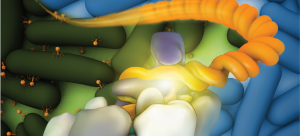
Structural impact of K63 ubiquitin on yeast translocating ribosomes under oxidative stress K63 ubiquitination of ribosomes serves as a key regulator of protein production during cellular exposure to oxidative stress. Defining the structural and functional mechanisms of translation regulation would support the current understanding of critical reprogramming of eukaryotic gene expression. Our paper presents an examination of the structure…
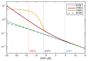
Mathematical Theory, Computational Challenges, and Opportunities Structural biology studies the structure and dynamics of macromolecules to broaden our knowledge about the mechanisms of life and impact the drug-discovery process. Owing to recent groundbreaking developments, chiefly in hardware technologies and data processing techniques, many new molecular structures have been elucidated to near-atomic resolutions using cryo-EM. The main goal of this article…
Disruption of the HIV-1 Envelope allosteric network blocks CD4-induced rearrangements The trimeric HIV-1 Envelope protein (Env) mediates viral-host cell fusion via a network of conformational transitions, with allosteric elements in each protomer orchestrating host receptor-induced exposure of the co-receptor binding site and fusion elements. To understand the molecular details of this allostery, here, we introduce Env mutations aimed to…
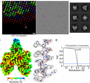
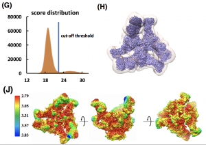
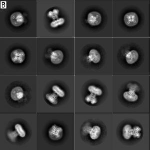
Unsupervised particle sorting for high-resolution single-particle cryo-EM Single-particle cryo-Electron Microscopy (EM) has become a popular technique for determining the structure of challenging biomolecules that are inaccessible to other technologies. Recent advances in automation, both in data collection and data processing, have significantly lowered the barrier for non-expert users to successfully execute the structure determination workflow. Many critical data processing…
Single-particle cryo-EM structure of a voltage-activated potassium channel in lipid nanodiscs The structure of a voltage-activated potassium channel in lipid nanodiscs solved using cryo-electron microscopy is similar to previous X-ray structures, and provides insights into the mechanism of C-type inactivation. eLife, 7:e37558, 2018.
Cryo-EM structure of human rhodopsin bound to an inhibitory G protein For the first time, scientists have visualized the interaction between two critical components of the body’s vast cellular communication network, a discovery that could lead to more effective medications with fewer side effects for conditions ranging from migraine to cancer. The near-atomic resolution images obtained, show a G-protein coupled…
Atomic Resolution Cryo-EM Structure of B-galactosidase We report methods to account for radiation damage and local changes in defocus and image drift, enabling visualization of atomic resolution features in a cryo-EM density map of inhibitor-bound b-galactosidase, and measuring of local flexibility of the bound inhibitor using constrained molecular dynamics simulations. Structure, 26(6), 2018.
Atomic Resolution Cryo-EM Structure of B-galactosidase We report methods to account for radiation damage and local changes in defocus and image drift, enabling visualization of atomic resolution features in a cryo-EM density map of inhibitor-bound -galactosidase, and derivation of atom-specific measures of local flexibility of the bound inhibitor using constrained molecular dynamics simulations. Structure, 26(6), p. 848-85, 2018.
Cryo-EM Structures Reveal Mechanism and Inhibition of DNA Targeting by a CRISPR-Cas Surveillance Complex Electron-microscopy images reveal how a CRISPR system marks specific DNA sequences for destruction. Microbes use CRISPR as a defense system to fend off viruses and other invaders, and geneticists have harnessed it to alter DNA sequences in a process called gene editing. We used cryo-electron microscopy…
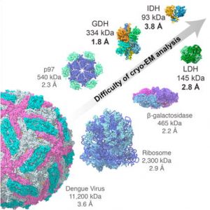
Breaking Cryo-EM Resolution Barriers to Facilitate Drug Discovery Using cryo-EM we were able to capture images of glutamate dehydrogenase (GDH), an enzyme found in cells, at a resolution of 1.8 angstroms, a level of detail at which the structure of the central parts of the enzyme could be visualized in atomic detail. We also imaged two small proteins in…