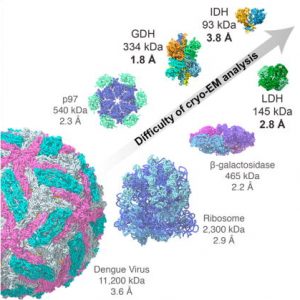
Breaking Cryo-EM Resolution Barriers to Facilitate Drug Discovery Using cryo-EM we were able to capture images of glutamate dehydrogenase (GDH), an enzyme found in cells, at a resolution of 1.8 angstroms, a level of detail at which the structure of the central parts of the enzyme could be visualized in atomic detail. We also imaged two small proteins in…
2.3 Å Resolution Cryo-EM Structure of Human p97 and Mechanism of Allosteric Inhibition The protein p97 is an AAA adenosine triphosphatase (ATPase) that uses energy from ATP hydrolysis to regulate substrates involved in intracellular protein quality control. Its role in this central process makes it a target for cancer chemotherapy. We used cryo-electron microscopy to determine high-resolution structures for…
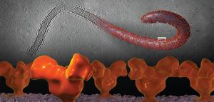
Cryo-electron Microscopy Structures of Chimeric Hemagglutinin Displayed on a Universal Influenza Vaccine Candidate Chimeric hemagglutinin proteins are set to undergo human clinical trials as a universal influenza vaccine candidate, yet no structural information for these proteins is available. Using cryo-electron tomography, we report the first three-dimensional (3D) visualization of chimeric hemagglutinin proteins displayed on the surface of the influenza…
Cryo-EM Structures of the Magnesium Channel CorA Reveal Symmetry Break upon Gating Magnesium ions (Mg2+) play essential roles in all living organisms. Bacteria and other prokaryotes rely upon the Mg2+-dependent channel CorA, which is composed of five identical subunits (A-E), to obtain these ions from their surroundings. Studies of CorA showed that, in contrast to most ligand-gated ion channels,…
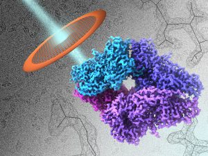
2.2 Å Resolution Cryo-EM Structure of β-galactosidase in Complex with a Cell-permeant Inhibitor Recent advances in cryo–electron microscopy allow structures of large macromolecules to be determined at near-atomic resolution. So far, though, resolutions approaching 2 Å, where features key to drug design are revealed, remain the territory of x-ray crystallography. Bartesaghi et al. achieved a resolution of 2.2 Å for a…
Structural Mechanism of Glutamate Receptor Activation and Desensitization Understanding the structural basis of the transition from closed to active and desensitized conformations is central to deciphering the function of ionotropic glutamate receptors NMDA receptors, AMPA receptors, delta receptors, and kainate receptors as mediators of excitatory synaptic transmission in the central nervous system. Ligand binding at the receptor's extracellular surface…
Structure of β-galactosidase at 3.2-Å Resolution Obtained by Cryo-Electron Microscopy The vast majority of high-resolution structures obtained using cryo-EM have been typically restricted to large, well-ordered entities such as helical or icosahedral assemblies or two-dimensional crystals. We show here that emerging methods in single-particle cryo-EM now allow structure determination at near-atomic resolution, even for much smaller protein complexes with…
Prefusion Structure of Trimeric HIV-1 Envelope Glycoprotein Determined by Cryo-Electron Microscopy HIV-1 Env transitions from a closed to an open state upon binding to its cellular receptor. Single-particle cryo-EM analysis now reveals the closed state of the HIV-1 Env trimer at ~6-Å resolution, featuring three gp41 helices at the center of the trimer. These findings indicate that HIV-1 enters…
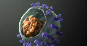
Molecular Architectures of Trimeric SIV and HIV-1 Envelope Glycoproteins on Intact Viruses: Strain-Dependent Variation in Quaternary Structure HIV and SIV contact and infect target T-cells following the binding of trimeric Env spikes displayed on the viral membrane with cellular receptors. The conformational changes in trimeric Env that are triggered by the interaction between trimeric Env and cell surface receptors…
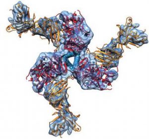
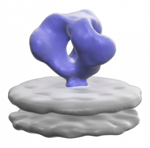
Molecular Architecture of Native HIV-1 gp120 Trimers We determined the structure of the native gp120 coat protein of HIV by cryo-electron tomography and molecular modelling. Comparison of gp120 structures in an unbound state, bound to a neutralizing antibody and bound to CD4 cell surface protein provides insight into the conformational changes that occur during antibody neutralization and attachment to…